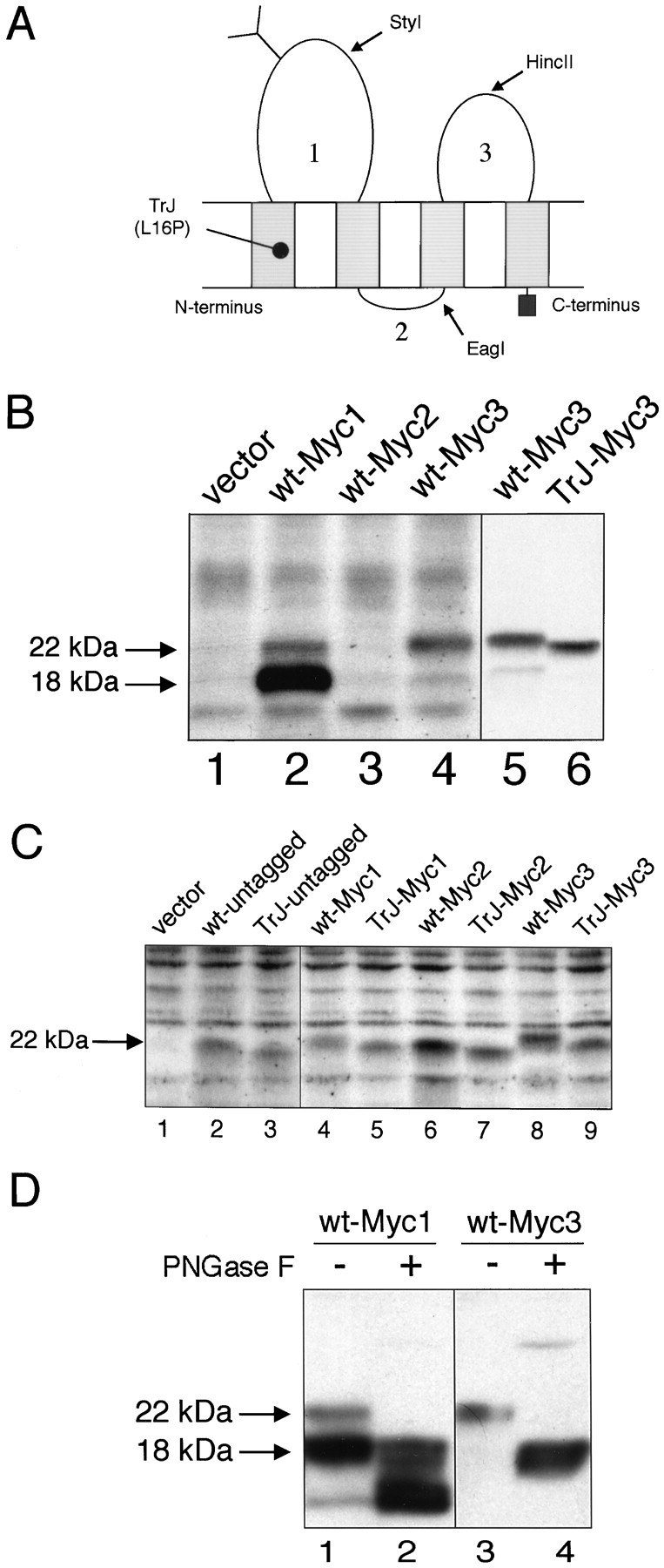
Fig. 1.

Characterization of the c-Myc epitope-tagged PMP22s. A, Expression of the c-Myc epitope in the three loops of PMP22. Shown is a schematic representation of PMP22. The position of the TrJ mutation is shown near the N terminus. The restriction sites used for inserting the tags are marked by arrows. Numbers represent the position of proposed loops 1, 2, or3. B, Immunodetection of c-Myc tagged wt- and TrJ-PMP22 in COS7 cells. The Myc epitope was inserted into loop 1 (Myc1), loop 2 (Myc2), or loop 3 (Myc3) of wt- or TrJ-PMP22. The expression vector without the cDNA insert was used as a control (vector). Cell lysates were prepared 36 hr after transfection and separated on 12.5% SDS gels; after blotting, the membranes were incubated with a polyclonal rabbit antibody against the c-Myc epitope. The positions of the arrows are derived from molecular weight markers that identify the N-glycosylated PMP22 (22 kDa) and the deglycosylated PMP22 (18 kDa). C, The expression of the c-Myc epitope in the three loops of wt- or TrJ-PMP22 monitored by an anti-PMP22 antibody. The Myc epitope was inserted into loop 1 (Myc1), loop 2 (Myc2), or loop 3 (Myc3) of wt- or TrJ-PMP22, as described in Materials and Methods. The expression vector without a cDNA insert (vector) was used as a control. Cell lysates were prepared 36 hr after transfection and separated on 12.5% SDS gels; after blotting, the membranes were analyzed with a polyclonal rabbit antibody against loop 3 of PMP22. The position of thearrow is derived from molecular weight markers and indicates the migration position of N-glycosylated PMP22 (22 kDa). D, Carbohydrate analysis of wt-Myc1 and wt-Myc3 shows the expected pattern for wt-Myc3, but not for wt-Myc1. Wt-PMP22 with the c-Myc epitope in loop 1 (Myc1) or loop 3 (Myc3) was expressed in COS7 cells. Cell extracts were prepared 36 hr after transfection, incubated with PNGaseF (+), and separated on 12.5% SDS gels. After blotting, the membranes were incubated with a polyclonal rabbit antibody against the c-Myc epitope. Untreated cell extracts were used as controls (−). The positions of the arrows are derived from molecular weight markers and indicate the position of N-glycosylated (22 kDa) and deglycosylated (18 kDa) forms of PMP22.
