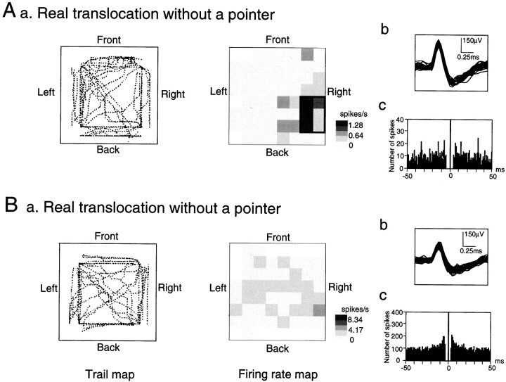
Fig. 3.
Two typical examples of the HF neurons.A, An example of an HF neuron with location-differential responses. Aa, The trail of the cab (left panel) and the corresponding location-differential responses (right panel) in the RT/TC task. A place field is surrounded by thick lines. Calibration is shown at the right; a mean firing rate for each pixel was expressed as a relative firing rate in which the mean firing rate in each pixel was divided by the grand mean firing rate in each task and was shown in five steps (R ≥ 2M; 2.0M > R ≥ 1.5M; 1.5M > R ≥ 1.0M; 1.0M > R ≥ 0.5M; R < 0.5M). Three values in the calibration indicate those of 2M, M, and 0, respectively. Regions not visited by the monkey during the session(s) are shown by blank pixels. Note that the activity increased in the right back corner of the experimental field, although the monkey moved and visited various sites of the experimental field and received juice reward at the four corners of the experimental field. Ab, Superimposed spike waves of the HF neuron shown in Aa. Ac, Autocorrelogram of the HF neuron shown in Aa. Ordinate, Number of spikes; abscissa, time in msec; bin size, 1 msec. Note that a refractory period of the neuron was 2–3 msec, which indicated that these spikes were recorded from a single neuron.B, An example of an HF neuron without location-differential responses. Note that no place fields were observed. Other descriptions as in A.

