
Fig. 6.
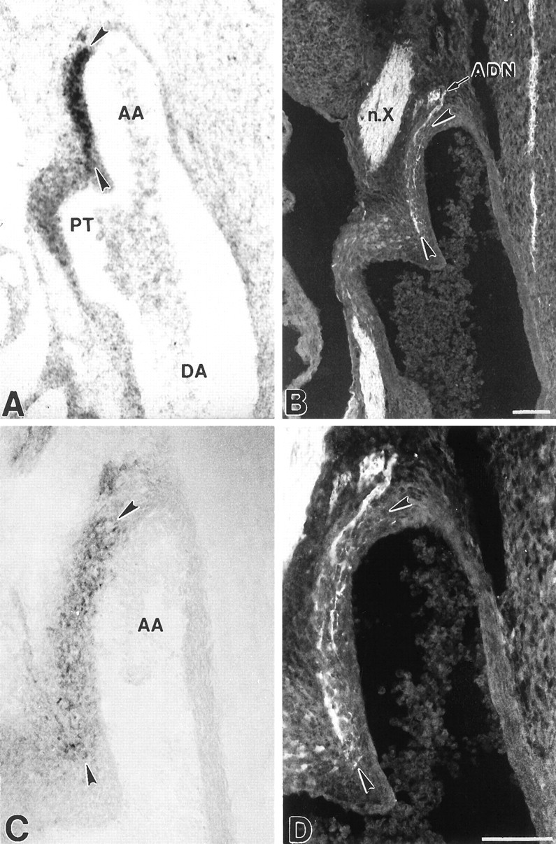
Localization of BDNF mRNA and protein, and baroreceptor fibers in the fetal aortic arch. Photomicrographs showing localization of BDNF mRNA (A), BDNF immunoreactivity (C), and PGP-stained baroreceptor fibers (B, D) in the E16.5 aortic arch (AA; arrowheads) at the level of the junction with the pulmonary trunk (PT). Note the correspondence between the distribution of baroreceptor fibers ramifying in the outer wall of the arch (B,arrowheads and at higher magnification inD) and BDNF mRNA and protein (A, C,arrowheads). ADN, Aortic depressor nerve;DA, descending aorta;n.X, vagus nerve. Scale bars, 100 μm.