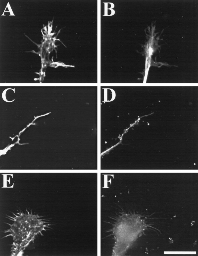
Fig. 3.
CNS myelin-associated growth inhibitors cause a rearrangement of actin filaments in motor neuron growth cones. Motor neurons were grown on FN for 2 d and then treated with PBS (A, B) or 100 μg/ml CNS myelin (C–F). Cultures were fixed and stained for actin filaments with rhodamine-labeled phalloidin (A, C, E) or for microtubules using a monoclonal anti-tubulin antibody followed by a fluorescein-conjugated secondary antibody (B, D, F). In controls, growth cones displayed many actin filament-rich filopodia and lamellipodia (A) with a dense bundle of microtubules defining the central region of the growth cones (B). On exposure to CNS myelin, growth cones retracted both lamellipodia and filopodia concomitant with a decrease in rhodamine fluorescence predominantly in the periphery.C, Often motor neuron growth cones responded to CNS myelin by retracting the entire peripheral region using the tubulin staining as our criteria. E, In some cases, motor neuron growth cones displayed stubby filopodial remnants with decreased rhodamine fluorescence in the peripheral region. Images were acquired using identical parameters. Scale bar, 10 μm.

