
Fig. 5.
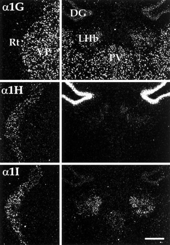
Distribution of CaVT expression in the thalamus. Left panels show differential labeling of the thalamic reticular nucleus (Rt) and ventral posterior thalamic nucleus (VP). Note that neurons of the reticular nucleus contained α1H and α1I mRNA, whereas α1G expression was limited to thalamic relay nuclei, including VP.Right panels show labeling in the habenulae and midline thalamic nuclei. Neurons of the lateral habenular nucleus (LHb) expressed α1G and α1I mRNA (see Results for details). DG, Dentate gyrus; PV, paraventricular thalamic nucleus. Scale bar, 250 μm.