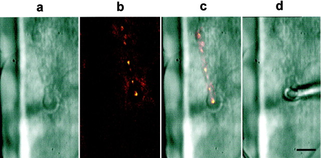
Fig. 2.
Macropatch recordings from individual, visualized synaptic boutons of the larval neuromuscular junction. Placement of the focal macropatch electrode over individual Ib boutons at the larval neuromuscular junction was under visual control to record synaptic currents generated at that site. a, Strings of Ib and Is boutons can be seen innervating the surface of muscle 6 under Nomarski optics. b, Under DiOC2(5) fluorescence, the same string of Ib boutons can be seen clearly, and the surrounding subsynaptic reticulum (SSR) does not fluoresce; however, fluorescence appears where the SSR meets the muscle. c, Overlay of the fluorescence image on the Nomarski image is shown.d, The focal macropatch electrode is gently placed over the chosen Ib bouton. The bouton can be seen through the lumen of the electrode. At this concentration of DiOC2(5) and exposure to illumination for fluorescence, synaptic transmission was unaffected compared with that in controls, and electron microscopy revealed no obvious damage to synaptic structure (S. Karunanithi, L. Marin, and H. L. Atwood, unpublished observations). Scale bar, 10 μm.

