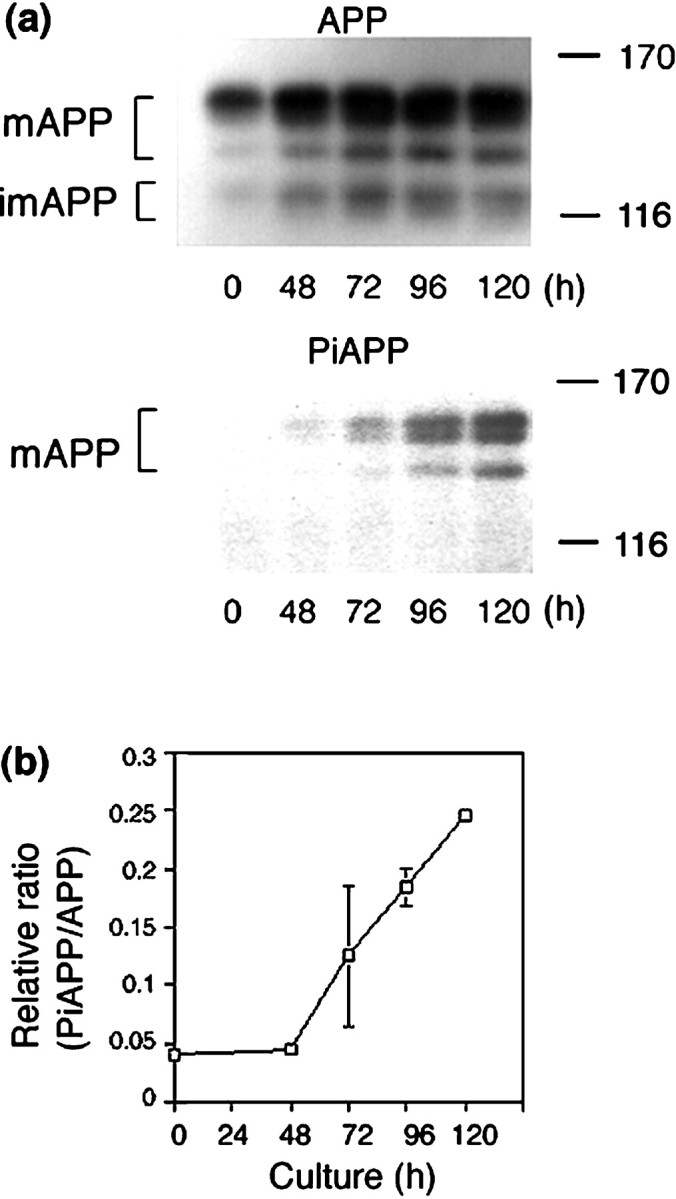
Fig. 1.

Phosphorylation of APP at Thr668 during neuronal differentiation of PC12 cells. PC12 cells (∼1 × 10⁶ cells) were cultured in the presence of NGF for the indicated times (0–120 hr). a, APP was immunoprecipitated from cell lysates (1.5 mg of protein) using UT-421, and samples were analyzed by SDS-PAGE [6% (w/v) polyacrylamide] and Western blotting using either UT-421 (APP,top) or UT-33 (PiAPP,bottom). b, APP and PiAPP were quantified using a Fuji BAS 2000 Imaging Analyzer, and the level of PiAPP was normalized to that of APP. The results shown are the average of duplicate assays, and error bars are indicated. mAPP, Mature APP isoforms; imAPP, immature APP isoforms. The size of the molecular weight standards (in kilodaltons) is indicated.
