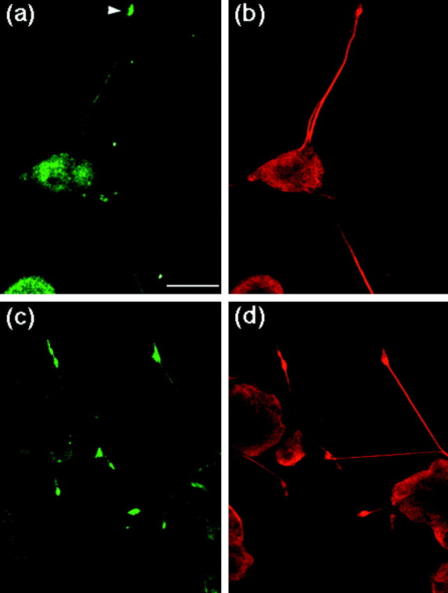
Fig. 2.

Localization of APP and phosphorylated APP in differentiating PC12 cells. PC12 cells ∼2–3 × 10⁴) were cultured for 72 hr with NGF. Cells were double-stained with UT-421 (a) and TU-01 (b) antibodies, or double-stained with UT-33 (c) and TU-01 (d). Scale bar, 25 μm. Arrowhead indicates a growth cone.
