
Fig. 3.
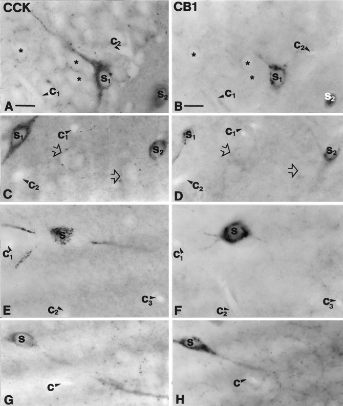
Cholecystokinin-containing interneurons express CB1 cannabinoid receptor in the hippocampus. A, B, A CCK-immunoreactive pyramidal-like basket cell (S1) in the dentate gyrus contains CB1-immunoreactivity. Granule cells (*) were negative for both markers.C, D, In the CA3 subfield large somata with thick proximal dendrites (S1) and smaller multipolar cells (S2) were positive for both CCK and CB1.Open arrows indicate double-negative cell bodies.E–H, These two morphological types colocalized CCK and CB1 in the CA1 subfield as well. A multipolar CCK-positive cell (S in E) in stratum radiatum is cut in half on the surface of the section. The same cell (S inF) shows CB1-immunoreactivity in the adjacent section. Three primary dendrites are also seen to continue in the adjacent section. G, H, A large bitufted neuron at the border of strata oriens and pyramidale is shown to be double-labeled. Capillaries labeled by c1–3 serve as landmarks to confirm precise alignment. CCK, Cholecystokinin;CB1, CB1 cannabinoid receptor. Scale bars (shown inA and B for A–H): 15 μm.