Fig. 2.

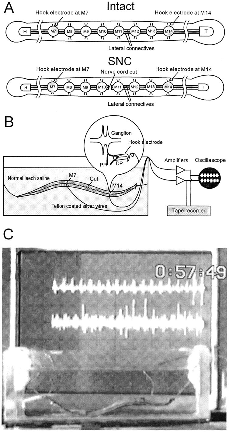
In situ recording.A, Two types of preparations used in the experiments. The leech ventral nerve cord is composed of a head ganglion (H), 21 midbody ganglia (M1–M21), and a tail ganglion (T). The median Faivre’s nerve and two lateral connective nerves link the ganglia. Interactions between the nerve cord and the body wall occur via the paired nerve roots that project from each midbody ganglion. The DP nerve, which branches from the posterior nerve root and innervates the dorsal side of the body wall, contains the large axon of the swim motoneuron DE-3.Top, Intact preparation with both the ventral nerve cord and the body wall intact. Bottom, SNC preparation with the ventral nerve cord severed between M10 and M11. B, Recording setup. Leeches were tethered by threads attached to head and tail suckers and suspended in a deep glass dish for physiological recording and videotaping. The lengths of threads tethering the leech were adjusted so that a full body wave could be developed. DP nerve activity was recorded in situ via fine silver hook electrodes. The inset illustrates the detail of a hook electrode. C, Snapshot of an experiment using the setup described above. The oscilloscope in the background displays signals recorded from the implanted electrodes.
