
Fig. 1.
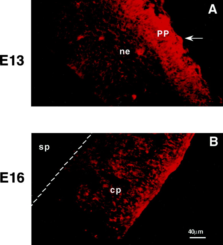
Glutamate immunoreactivity in the embryonic murine cortex. Photomicrographs of coronal sections of the embryonic cortex immunostained with anti-glutamate antisera.A, At E13, glutamate-immunoreactive cells (arrow) and processes are evident in the primordial plexiform layer (PP). B, By E16, fibers in the outer half of the cortical plate are highly immunoreactive for glutamate. ne, Neuroepithelium; cp, cortical plate; sp, subplate. Scale bar, 40 μm.