Fig. 1.
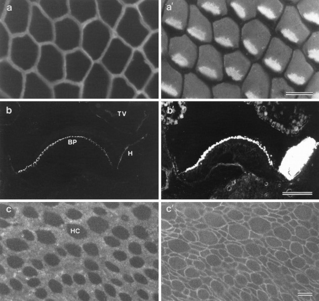
a, a′, A whole-mount preparation of the basilar papilla double-labeled with mAb D37 (a) and monoclonal anti-hair cell antigen (a′). The image is focused on the apical surface of the epithelium, in an area from the inferior region of the papilla where the short hair cells are located. MAb D37 stains the narrow compacted surfaces of the supporting cells that surround each hair cell. The anti-HCA mAb stains the base of the hair bundle and most of the apical, nonstereociliary surface of the cell except for a small patch lying behind the hair bundle where the kinocilium is located. b, b′, A cryosection of the basilar papilla double-labeled with mAb D37 (b) and rhodamine–phalloidin (b′) to reveal the distribution of the SCA and F-actin, respectively. Note how the surface of the basilar papilla (BP) is intensely stained by mAb D37. Staining is also observed on the surfaces of the homogene cells (H). The lumenal surface of the tegmentum vasculosum (TV) is only very weakly labeled.c, c′, A whole-mount preparation of the utricular macula double-labeled with mAb D37 to reveal the distribution of the SCA (c) and rabbit anti-cingulin to distinguish the boundaries of the cells (c′). The apical surfaces of the hair cells (HC) are not labeled by mAb D37 and appear as dark holes (c). Scale bars: a,a′, c, c′, 10 μm;b, b′, 100 μm.

