
Fig. 10.
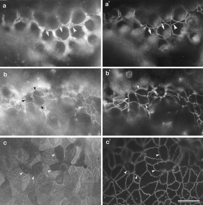
In vitro preparations of a control basilar papilla (a, a′) and basilar papillae that have been treated with either 1.0 mm (b, b′) or 0.2 mm (c, c′) neomycin for 24 hr and triple-labeled to visualize the SCA through one channel (a–c), and both cingulin and the HCA simultaneously through the other channel (a′–c′). a, a′, In control papillae, strong staining for the SCA is seen on the supporting cells (a) that surround the HCA-positive hair cells (a′). Arrowheadsin a and a′ point to the same supporting cell; arrows point to the same hair cells. b, b′, c, c′, In neomycin-treated papillae, SCA-negative cells are seen that are in contact with each other (arrowheads). These cells are also HCA-negative and do not have the circular apical surfaces typical of hair cells (a′). Scale bar (shown inc′ and applies to all panels): 10 μm.