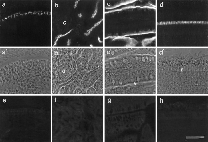
Fig. 3.
Immunofluorescence micrographs (a–d) and the corresponding phase-contrast images (a′–d′) of cryosections from the basilar papilla (a), kidney (b), intestine (c), and retina (d) stained with mAb D37. In a, the surfaces of the supporting cells in the basilar papilla give a honeycomb staining pattern. Inb, the brush borders of tubules in the kidney are stained but the glomerulus (G) is unstained. Inc, the brush borders of the intestinal villi (V) are stained, and in d, the external limiting membrane (E) marking the boundary of the outer nuclear layer is stained. e–h, Immunofluorescence micrographs of cryosections of basilar papilla (e), kidney (f), intestine (g), and retina (h) stained with an irrelevant IgG2bmAb, MOPC 141. Scale bar, 50 μm (applies toa–h).

