
Fig. 9.
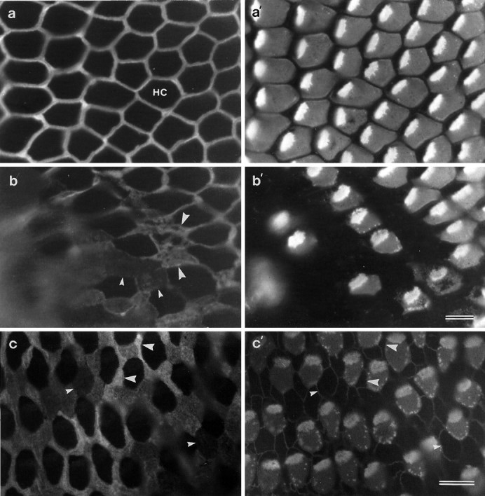
Whole-mount preparations from noise-damaged basilar papillae that have been double-labeled with mAb D37 (a, b) and monoclonal anti-HCA (a′, b′), or triple-labeled with mAb D37 (c) and a mixture of monoclonal anti-HCA and cingulin (c′). Images ina and b are from a papilla exposed to octave band noise centered around 2.0 kHz at 120 dB SPL for 14 hr; the image in c is from a papilla exposed to a 900 Hz tone at 120 dB SPL for 12 hr. a, a′, Area of the inferior basilar papilla just outside the noise-damaged region, showing narrow supporting-cell apical surfaces strongly labeled for the SCA (a) and structurally normal, HCA-positive hair cells (a′). b, b′, Edge of the noise-damaged region showing hair cells with varying degrees of damage. Note that supporting cells further into the damaged region express less SCA (small arrowheads) than supporting cells at the edge of the damaged region (large arrowheads). c, c′, Region of a noise-damaged basilar papilla showing supporting-cell surfaces (c) that label strongly (large arrowheads) or very weakly for the SCA (small arrowheads). Note the speckled appearance of the HCA staining around the perimeter of the noise-damaged hair cells (b′, c′). Scale bar (shown in b′):a–b′, 10 μm; (shown in c′): c, c′, 10 μm.