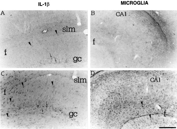
Fig. 4.

High-magnification photomicrographs showing IL-1β immunoreactivity (A, C) and B4-isolectin-positive microglia (B, D) in coronal sections of the rat dorsal hippocampus 24 hr after a local injection of PBS (A, B) or 0.19 nmol of kainic acid (C, D). Note the diffuse pattern of enhanced IL-1β immunoreactivity in glia-like cells (C) and the widespread staining of B4-isolectin-positive microglia (D). Scattered IL-1β immunoreactive neurons were also observed (A, C, arrows). gc, Granule cells; f, fissura hippocampi;CA1, CA1 pyramidal layer; slm, Stratum lacunosum moleculare. Scale bar, 200 μm.
