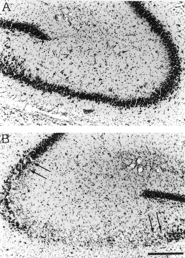
Fig. 5.

Nissl staining of coronal sections of the dorsal hippocampus of a representative rat 1 week after the injection of PBS (A) or 0.19 nmol of kainic acid (B). Note the loss of CA3 neurons (B, arrows) that was restricted to the injected dorsal hippocampus. Scale bar, 200 μm.
