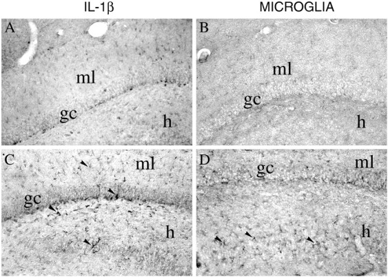
Fig. 6.

Photomicrographs showing IL-1β immunoreactivity (A, C) and B4-isolectin-positive microglia (B, D) in coronal sections of the rat dorsal hippocampus 3 hr after a local injection of 12.5% PEG (A, B) or 0.77 nmol of bicuculline methiodide (C, D). IL-1β immunoreactivity was markedly increased in glia-like cells located in the granule cell layer and in the molecular layer (ml) of the dentate gyrus (arrowheads). These cells have a darkly stained cell body and branched processes. Some of these cells were in close proximity to granule cells (gc) and interposed between CA3c neurons in the hilus (h;C, arrowheads). B4-isolectin-positive microglia was increased in the same regions (D). Scale bar, 200 μm.
