Fig. 3.
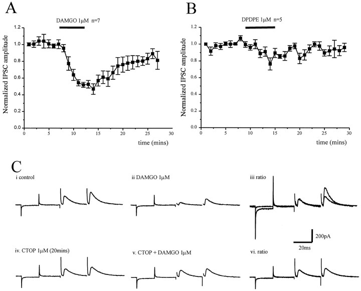
Presynaptic depression of evoked striatopallidal IPSCs by μ opioid receptor activation. A, Time course showing the effect of the μ opioid receptor agonist DAMGO (1 μm) on the normalized IPSC amplitude evoked by stimulation of striatum in parasagittal slices in the presence of CNQX (10 μm) and dl-AP-5 (100 μm). DAMGO reduces the IPSC by 53 ± 6% (n = 7).B, Time course showing the effect of the δ opioid receptor agonist DPDPE (1 μm) on the normalized IPSC amplitude evoked by stimulation of the striatum in parasagittal slices in the presence of CNQX (10 μm) and dl-AP-5 (100 μm). DPDPE has no significant effect on the evoked IPSC (n = 5). However, the IPSC evoked in each of these five cells was inhibited by DAMGO (1 μm).C, Paired IPSCs recorded from the same cell in response to the same stimulation at an interval of 50 msec in control (i) and DAMGO (1 μm) (ii). iii, The pair of IPSCs recorded in the presence of DAMGO have been scaled so that the conditioning (first) responses with and without drug are of similar amplitude. The ratio of the test (second) response in DAMGO has increased, indicative of a presynaptic site of action. Paired IPSCs recorded in the presence of the μ opioid receptor antagonist CTOP (1 μm) (iv) and CTOP plus DAMGO (1 μm) (v), and ratio (vi). In the presence of CTOP, there was no depression of the conditioning IPSC by DAMGO and no change in the paired-pulse ratio. Currents resulting from a 10 mV hyperpolarizing step precede all IPSC pairs.

