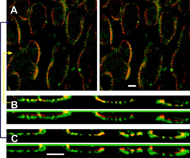
Fig. 4.
Confocal imaging of synaptic membrane and postsynaptic folds. Deconvolved images show light-level colocalization of the basal lamina-specific lectin FITC-VVA (green; synaptic cleft) and the anti-AChR monoclonal mAb22 (red; postsynaptic membrane).A, Stereo view from above, looking through a small region of one nerve terminal toward the muscle below. Note thepeanut-shell shapes of boutons’ invaginations into muscle fiber; this curved surface also identifies the presynaptic membrane within light resolution.Fingerprint-like stripes are postsynaptic folds, which can be seen to radiate into the muscle fiber at the edges of boutons. B, Top, Magnifiedx–z cross section at the yellow arrow in A. Bottom,Red and green images displaced vertically so that the labels can be viewed individually. C, Magnified x–z views as inB taken at the blue arrow inA. Folds are visible and nearly coincident with both labels. Scale bars, 2 μm.

