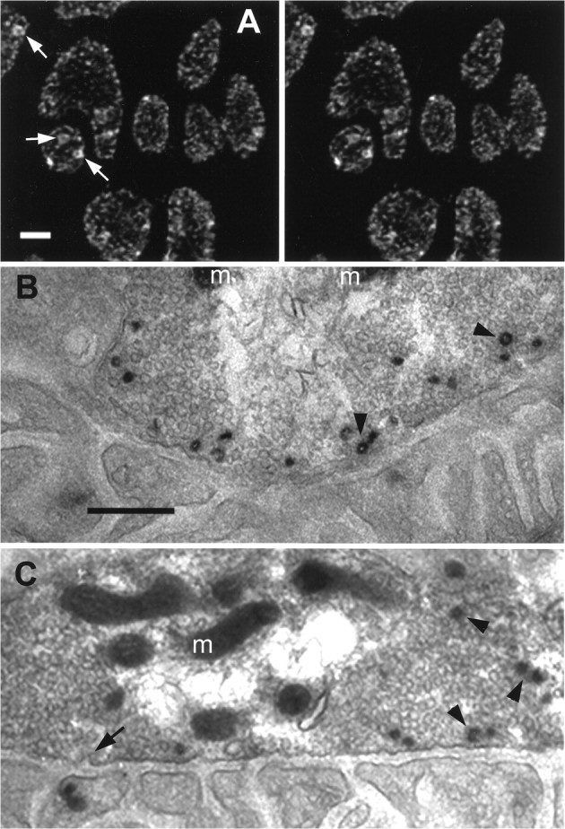
Fig. 8.

Labeled structures were probably vesicle clusters and endosomes. A, Uptake of the membrane-permeant dye FM1-43 was similar to that of the aqueous dye SR101 but permitted photoconversion for EM. Shown is the staining pattern (stereo view) obtained after brief stimulation (5 Hz; 40 sec) at ∼7°C and fixation as described in Materials and Methods (compare with Fig.2D). Large structures often appeared hollow (arrows), suggesting that they were bound by a membrane and were not clusters of vesicles. B, C, Shown are example EMs of photoconverted FM1-43 from two boutons, as inA but with briefer stimulation (5 Hz; 30 sec) at ∼7°C so that most or all of the internalized dye appeared assmall dots.Sections shown are nearly tangential to the muscle fiber surface and close to the region of the bouton’s deepest invagination. The postsynaptic membrane and its folds arebelow, with the vesicle-filled boutonabove (m, mitochondria). Vesicles containing recently internalized FM1-43 were almost exclusively near the presynaptic membrane, the same location as the small dots seen at light level. Putative endocytic profiles (arrow) appeared in the presynaptic membrane. Some labeled vesicles were coated (arrowheads) as were some endocytic profiles (arrow). Scale bars:A, 2 μm; B, C, 0.5 μm.
