
Fig. 3.
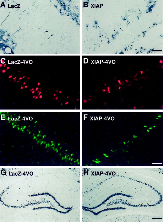
Attenuation of ischemia-induced caspase-3 activation and apoptotic death in CA1 neurons by XIAP overexpression. Animals received intrahippocampal injections of adenoviral constructs containing either lacZ (left side) or XIAP (right side) and were subjected to 4-VO or sham surgery 5 d later. Virally mediated overexpression of lacZ and XIAP was confirmed by immunohistochemical detection of β-galactosidase and human XIAP, respectively. The majority of immunoreactive cells in the CA1 layer appeared to be pyramidal neurons. However, a small number of glial cells were also infected with the viral constructs. Scale bar (in B):A, B, 75 μm. In animals that received intrahippocampal injections of the adenoviral constructs followed by sham 4-VO (n = 4), approximately the same number of CA1 neurons were immunoreactive for β-galactosidase (9 ± 2) and human XIAP (10 ± 3). Levels of catalytically active caspase-3 were assessed in CA1 neurons by immunohistochemical detection of the (p17/p12)2 tetramer 48 hr after 4-VO (n= 4) (C, D). In the hippocampus injected with the lacZ construct, a large number of neurons were stained in the medial aspect of CA1 (C). In contrast, very few immunoreactive neurons were present in the contralateral hippocampus that had been injected with the XIAP construct (D). DNA fragmentation was examined 5 d after 4-VO in animals injected with these viral constructs. A larger number of ISEL-positive CA1 neurons were observed in the lacZ-injected side (E). In contrast, considerably fewer ISEL-positive neurons were detected in the XIAP-injected side (F). Scale bar (in F):C–F, 75 μm. Immunohistochemistry for NeuN was performed to stain surviving neurons 7 d after 4-VO (12 min). A small number of neurons was observed in the CA1 subfield injected with the lacZ construct (G), whereas the majority of neurons in the XIAP-injected side remained NeuN-positive (H). Scale bar (inH): G, H, 400 μm.