Fig. 1.

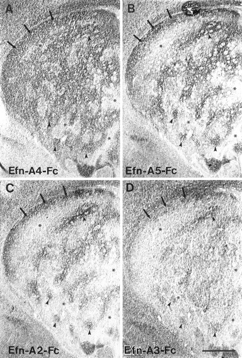
Adjacent coronal sections through the striatum of a P6 rat incubated with four different ephrin-A–Fc fusion proteins. A, Ephrin-A4–Fc (Efn-A4–Fc) displays extensive binding to large areas of the striatal neuropil (arrowheads) that are perforated by smaller regions exhibiting less dense binding (asterisks). A prominent continuous region of binding is consistently observed along the dorsal aspect of the striatum (arrows).B, C, Ephrin-A5–Fc (Efn-A5–Fc) (B) and ephrin-A2–Fc (Efn-A2–Fc) (C) exhibit an identical binding pattern within the striatum, which differs from that observed for ephrin-A4–Fc (A). Both ephrin-A2–Fc and ephrin-A5–Fc exhibit low levels of binding to large areas of the striatum (asterisks). These areas are superimposed on the smaller regions that lack ephrin-A4–Fc binding (asterisks). As with ephrin-A4–Fc, these ligands exhibit a dense band of binding along the dorsal aspect of the striatum (arrows). Furthermore, these ligands exhibit more restrictive binding to areas of striatal neuropil (arrowheads) that are located within regions exhibiting ephrin-A4–Fc binding. D, The ephrin-A3–Fc (Efn-A3–Fc) binding pattern is most similar to that observed for ephrin-A2–Fc and ephrin-A5–Fc, although there is less noticeable differentiation between striatal areas with high (arrows and arrowheads) versus low binding (asterisks). Scale bar, 500 μm.
