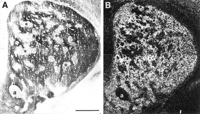
Fig. 5.

Comparison of ephrin-A4–Fc binding and EphA4 mRNA expression in P6 striatum. Adjacent coronal sections were incubated with ephrin-A4–Fc fusion protein (A) or processed for in situ hybridization for EphA4 mRNA (B). A, Bright-field micrograph showing the binding pattern of ephrin-A4–Fc. B, Dark-field micrograph showing in situ hybridization of EphA4 mRNA. Asterisks indicate corresponding areas between the two adjacent sections that lack ephrin-A4–Fc binding and EphA4 message. a, Anterior commissure. Scale bar, 500 μm.
