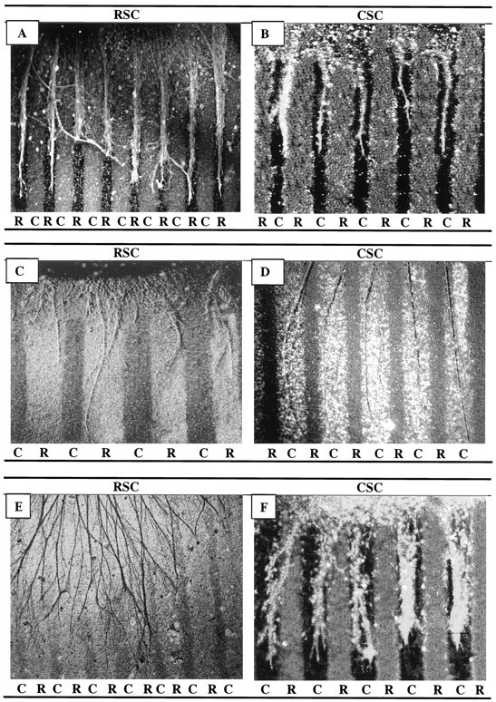
Fig. 1.

Position-dependent preferential outgrowth of spinal neurites on membranes from P0 and E18 limb muscles. Spinal cord slices cut from the cervical (rostral, RSC) or lumbar (caudal, CSC) enlargement were placed on alternating stripes of membrane derived from forelimb (rostral) or hindlimb (caudal) muscles. Membranes applied to one set of lanes in each culture were mixed with fluorescein-labeled beads to mark lane boundaries and marked rostral (R) or caudal (C). After 3 d, the cultures were fixed, and neurites were labeled with antibodies to neurofilaments (A, B), left unstained (C, D), or reacted with HRP-DAB(E) (see Materials and Methods). Rostral spinal cord neurites grew selectively on P0 rostral membranes in 31% of cultures(A) and on E18 rostral membranes in 9% of cultures(C). Caudal spinal cord neurites grew selectively on caudal P0 membranes in 52% of cultures (B) and on caudal E18 membranes in 78% of cultures (D). E, Rostral neurites grew randomly in 42% of cultures on E18 rostral membranes. F, Neurites of caudal spinal cord growing on caudal P0 membranes and stained with fluorescein-labeled antibody to acetylcholine transferase. Magnification: A, E, 60×;B, D, F, 100×; C, 140×.
