
Fig. 1.
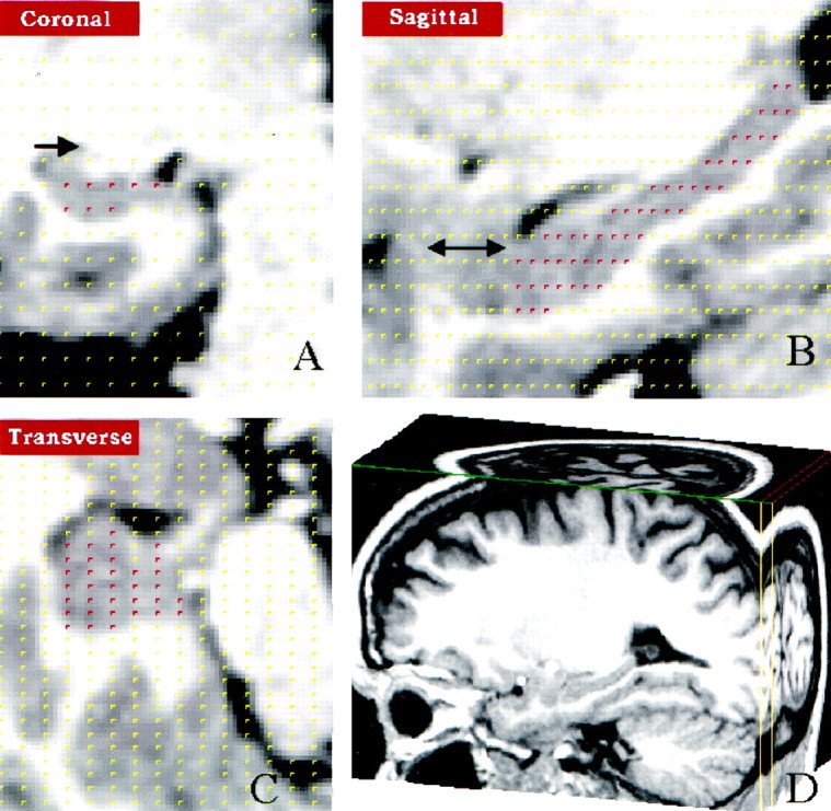
Hippocampal volume measurement using stereology.A, Coronal section through the hippocampus. Points within the hippocampus highlighted in red were selected from a randomly placed 5 × 5 mm2 grid overlying the hippocampus and are simultaneously displayed in (A) coronal, (B) sagittal, and (C) horizontal views. Arrowindicates the caudate (A), and ↔ indicates the amygdala 171 (B). D, Cubic volumes containing the hippocampus were sectioned out from the total brain volume.