
Fig. 10.
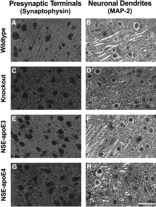
Differential effects of apoE3 and apoE4 on neuronal integrity in untreatedApoe−/− mice. Sections of neocortex from 7- to 9-month-old wild-type (A, B),Apoe−/− (C, D), NSE-apoE3 (E, F), and NSE-apoE4 (G, H) mice were immunostained for synaptophysin (A, C, E, G) or for MAP-2 (B, D, F, H) and imaged by confocal microscopy. Cases with severe damage were selected for illustration. Note the prominent loss of immunolabeled neuronal structures in the neocortex ofApoe−/− mice and NSE-apoE4 mice and the normal appearance of corresponding sections from wild-type and NSE-apoE3 mice. Qualitatively similar results were obtained for synaptophysin-positive presynaptic terminals in the hippocampus (data not shown). Scale bar, 55 μm.