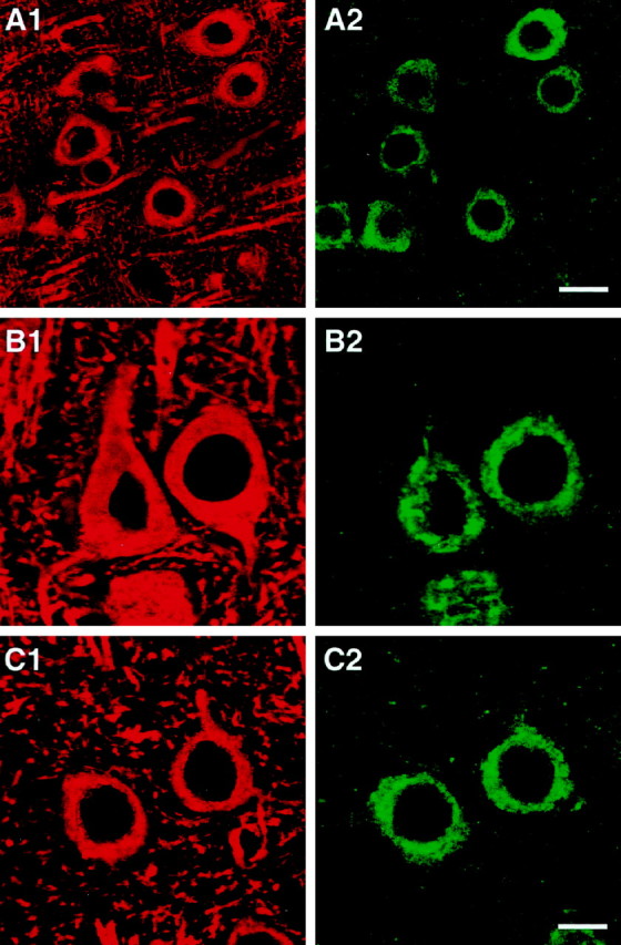
Fig. 6.

Colabeling of cells in NSE-apoE mouse brains for human apoE and for the neuronal marker MAP-2. Brain sections were double-immunolabeled with antibodies against human apoE (green; A2, B2, C2) and antibodies against MAP-2 (red; A1, B1, C1) and imaged by confocal microscopy. Pseudocolored images depict colabeled neurons in neocortex of an NSE-apoE3 (A, B) and of an NSE-apoE4 (C) mouse. Scale bars: A1, A2, 15 μm; B1, B2, C1, C2, 7 μm.
