
Fig. 2.
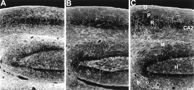
Dark-field photomicrograph, sagittal plane, of 5-HT immunoreactive axons in the hippocampus and dentate gyrus of a control monkey (A), a monkey treated with MDMA 2 weeks previously (B), and a monkey treated with MDMA 7 years previously (C). In both MDMA-treated monkeys, a reduction in axon density is evident in the stratum oriens (O), the stratum pyramidale (P), and the molecular layer (M). Note substantial denervation in the hilus (H) of the 7-year MDMA monkey. Scale bar, 200 μm.