
Fig. 4.
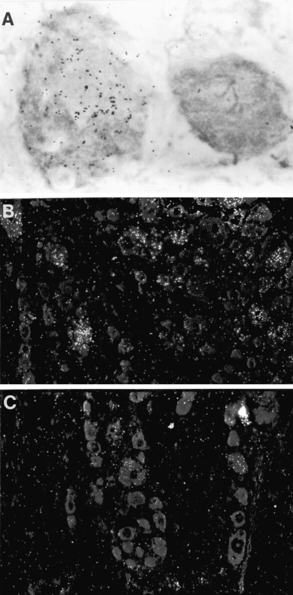
A, Light-field photomicrograph of IL-6 in situ hybridization preparation of L5 DRG ipsilateral to a sciatic nerve that had been injected with 48/80, a mast cell degranulating agent. One heavily labeled neuron is seen.B, Dark-field photomicrograph of IL-6 in situ hybridization preparation of DRG ipsilateral to transected sciatic nerve. C, Dark-field photomicrograph of IL-6in situ hybridization of DRG ipsilateral to sciatic nerve transection in a rat that had also been injected intraperitoneally with a mast cell stabilizing agent, cromolyn sodium. Note that the labeling evident after nerve transection is reduced by injection of cromolyn sodium. Magnification: A, 1120×;B, C, 180×.