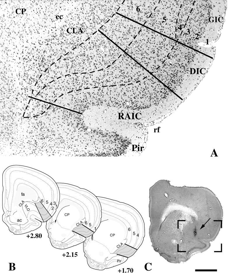
Fig. 1.

Landmarks of the RAIC that were used to define the “on-site” injections. A, Low power of a Nissl-stained transverse brain section through the middle third of the RAIC. The dorsal and ventral borders of the RAIC are delineated by thecontinuous bottom two lines. The RAIC’s ventral border extends laterally from the medial part of the rhinal fissure (rf) to the ventral tip of the claustrum (CLA). This ventral border marks the end of the four-layer transitional cortex, which becomes the three-layered piriform cortex (Pir). Dorsally, the border between the RAIC and the dysgranular insular cortex (DIC) is a line that extends perpendicularly from the cortical surface to the junction between the middle and dorsal third of the claustrum. Although a nascent lamina 4 is recognized in the differential interference contrast, it is well developed dorsally and serves to mark the border (top full line) with the granular insular cortex (GIC). The cortical layers are indicated inarabic numerals. B, Diagrams of transverse brain sections modified from the atlas of Swanson (1992). The gray areas between continuous lines represent the RAIC. Laminae 1–6 are delineated by interrupted lines. The rostrocaudal levels of each section according to Swanson’s atlas are indicated under each section. C, Whole transverse section of the right hemisphere, showing the injection site of the D1 receptor antagonist SCH-23390 (arrow). The boxed area delineates an area equivalent to that included inA. This animal displayed clear hyperalgesia to nociceptive heat in addition to allodynia to light touch.ac, Anterior commissure; CP, caudate-putamen; ec, external capsule;fa, anterior forceps of the corpus callosum;VLO, ventrolateral orbital cortex. Scale bar (shown inB): A, 200 μm; B, 1.7 mm.
