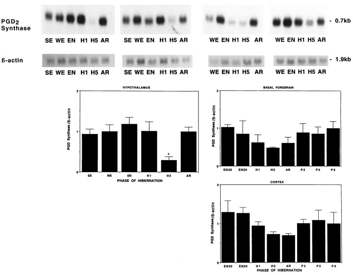
Fig. 8.
PGD2 synthase mRNA declines during torpor. Autoradiographs of Northern blots of total RNA (20 μg/lane) isolated from hypothalamus along with graphic representations of hypothalamus, cerebral cortex, and basal forebrain of ground squirrels killed during different phases of hibernation cycle and hybridized to a [32P]-labeled PGD2 synthase cDNA probe. Note the decreased level of PGD2 synthase mRNA during late torpor in the hypothalamus relative to all other conditions; *p< 0.05. The lower levels of PGD2 synthase mRNA in the cortex and basal forebrain during late torpor and arousal do not quite reach statistical significance. Filters for the hypothalamus are the same as used previously, and the cortical and basal forebrain samples are from the same animals as the hypothalamus shown in Figure 6.

