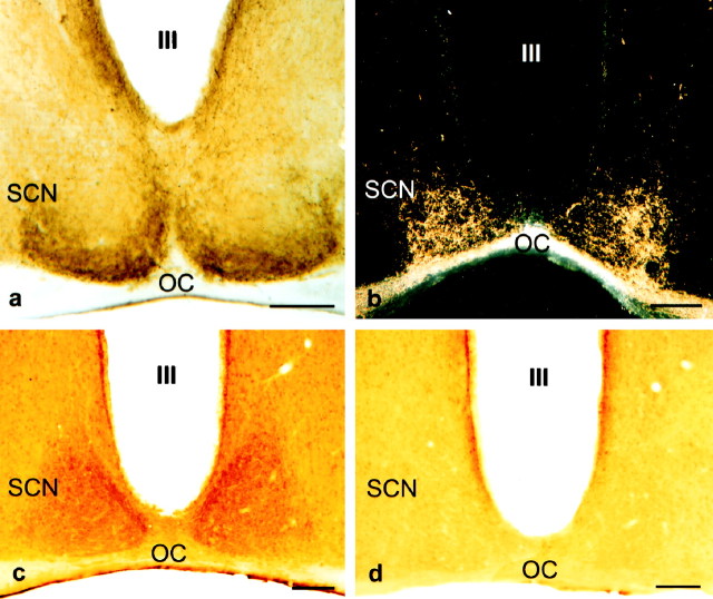
Fig. 1.
Light micrographs of coronal sections through the mouse anterior hypothalamus illustrating the 5-HT-immunoreactive process in the mid-caudal SCN (a), the HRP-labeled retinal processes in the mid-caudal SCN viewed using dark-field optics (b), 5-HT1Breceptor immunoreactivity in the mid-caudal SCN (c), and the absence of 5-HT1Breceptor immunoreactivity in the SCN treated with 5-HT1Breceptor antiserum preabsorbed with the peptide used for raising the antiserum (d). OC, Optic chiasm;III, third ventricle. Scale bars, 100 μm.

