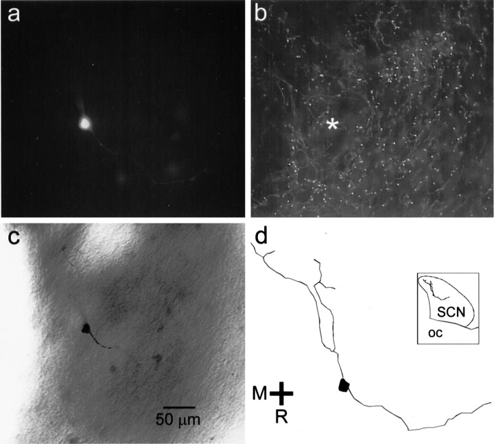
Fig. 7.
Location of an SCN neuron that responded to optic nerve stimulation and to TFMPP. a, This SCN neuron was filled with biocytin during a whole-cell patch-clamp recording and visualized with an avidin–rhodamine conjugate. b, The same tissue was labeled for 5-HT immunoreactivity. Immunopositive fibers are present near the position of the recorded neuron (asterisk). c, The same neuron was rereacted with avidin–biotin–horseradish peroxidase complex and visualized with DAB. d, The neuron was reconstructed digitally using Neurolucida (MicroBrightField). Inset, The position of the neuron relative to the SCN borders in thehorizontal plane of view is shown. OC, Optic chiasm.

