
Fig. 3.
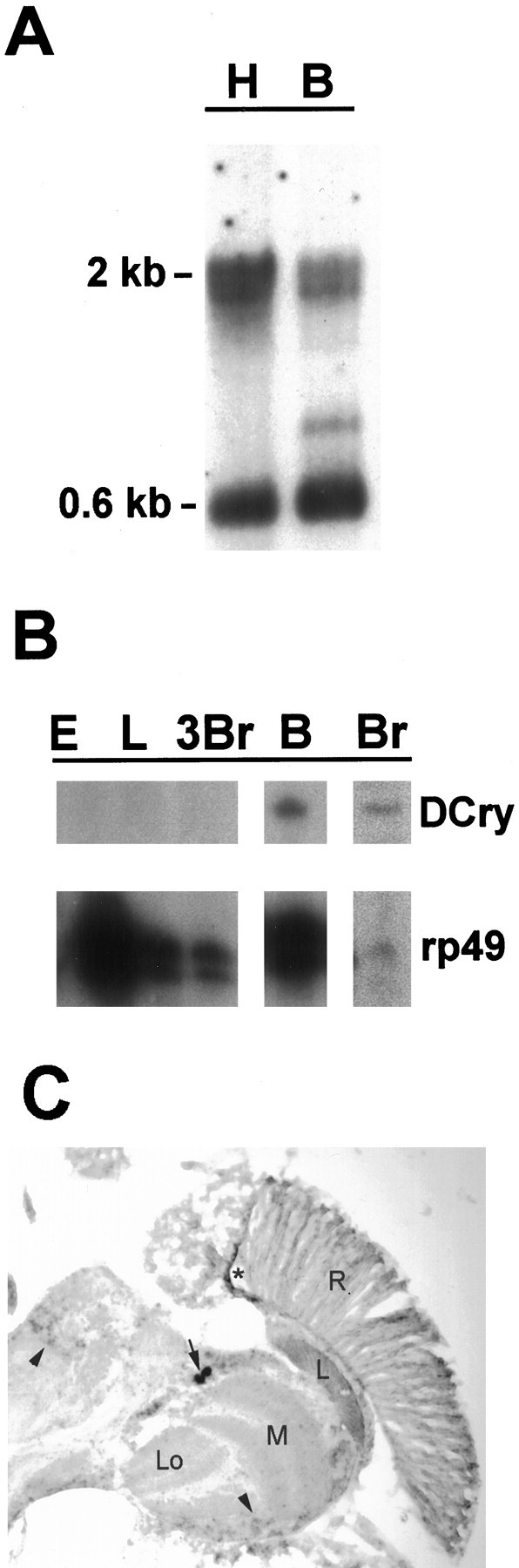
Tissue and developmental expression ofDCry mRNA. A, Northern analysis showing expression in head and body tissues. H denotes 5 μg of head poly(A+) RNA; B denotes 5 μg of body poly(A+) RNA. Size markers indicate theDCry mRNA doublet (2 kb) andrp49 mRNA (0.6 kb). B, RNase protection analysis of DCry expression in different tissues and developmental stages. Protected bands representing DCry and control rp49 mRNAs are indicated. Br, Adult brain; E, 0–24 hr embryo; L, third instar larva; 3Br, third instar larval brain; B, body. The body preparation used for RNase protection analysis was examined by light microscopy to be certain that no contaminating heads were present. C,In situ hybridization to a horizontal section of an adult head. The arrow indicates the position ofDCry-expressing cells in the lateral CNS. Thearrowheads show specific signal in other portions of the brain. The star indicates nonspecific staining, which also was seen with the sense probe. A similar spatial pattern of expression was observed in two independent experiments.R, Retina; L, optic lamina;M, optic medulla; Lo, lobula.