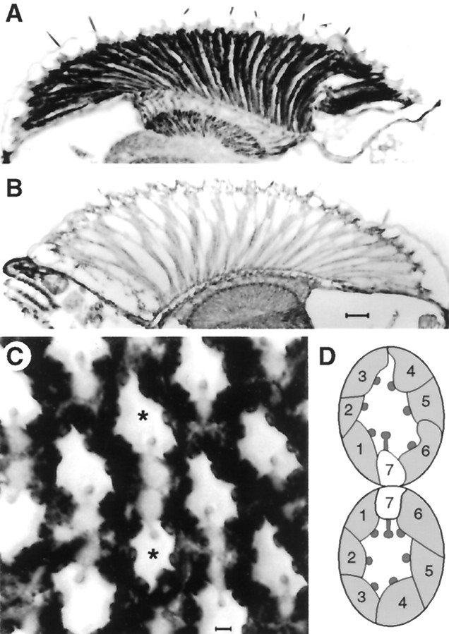
Fig. 3.
Frozen sections of fly heads stained with antibodies for Drosophila Gsα.A, Transverse section through Gsα* flies showing high levels of Gs in the photoreceptor cell bodies and axon terminals in the lamina compared with control (B) untransformed flies. Scale bar, 20 μm.C, Tangential section through a Gsα* eye at the equator showing high levels of immunoreactivity in cell bodies of photoreceptors R1–R6 compared with the central cell R7. Scale bar, 2 μm. D, A schematic diagram of the twoasterisked ommatidia indicating photoreceptors 1–7 (8 is below the plane of section) within each unit. Note that the two ommatidia straddle the equator of the eye and so are mirror images of each other.

