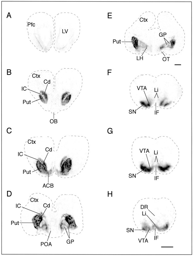
Fig. 3.
DAT binding sites are highly expressed in day 70 fetal monkey brain. Bright-field view of autoradiograms illustrating the distribution of DAT binding sites, labeled with [125I]RTI-121, in coronal sections from rostral (A) to caudal (H). The darker the images, the denser are the binding sites.A, The DAT binding sites were faint (0.08 ± 0.03 nCi/mg) in the prefrontal cortex (Pfc) and were illustrated within the intermediate layer after 3 weeks of film exposure. B–E, Within the caudate (Cd) and the putamen (Put) the DAT binding sites were dense and patchy in appearance, similar to the TH fiber stain illustrated in Figure 1. DAT binding sites were also present in the nucleus accumbens (ACB), globus pallidus (GP), and lateral hypothalamus (LH) and exhibited a distribution pattern similar to IR-TH. F–H, The highest density of DAT binding sites was measured in the substantia nigra (SN) and the ventral tegmental area (VTA) of the fetal midbrain. F–H, Light binding densities were found in the linear nucleus (Li) and interfascicular nucleus (IF), whereas binding sites were not detected in the dorsal Raphé (DR). The nonspecific binding is 0.04 ± 0.01 nCi/mg.Ctx, Cortex; IC, internal capsule;OB, olfactory bulb; OT, optic tract;POA, preoptic area. Scale bar, 2 mm.

