
Fig. 2.
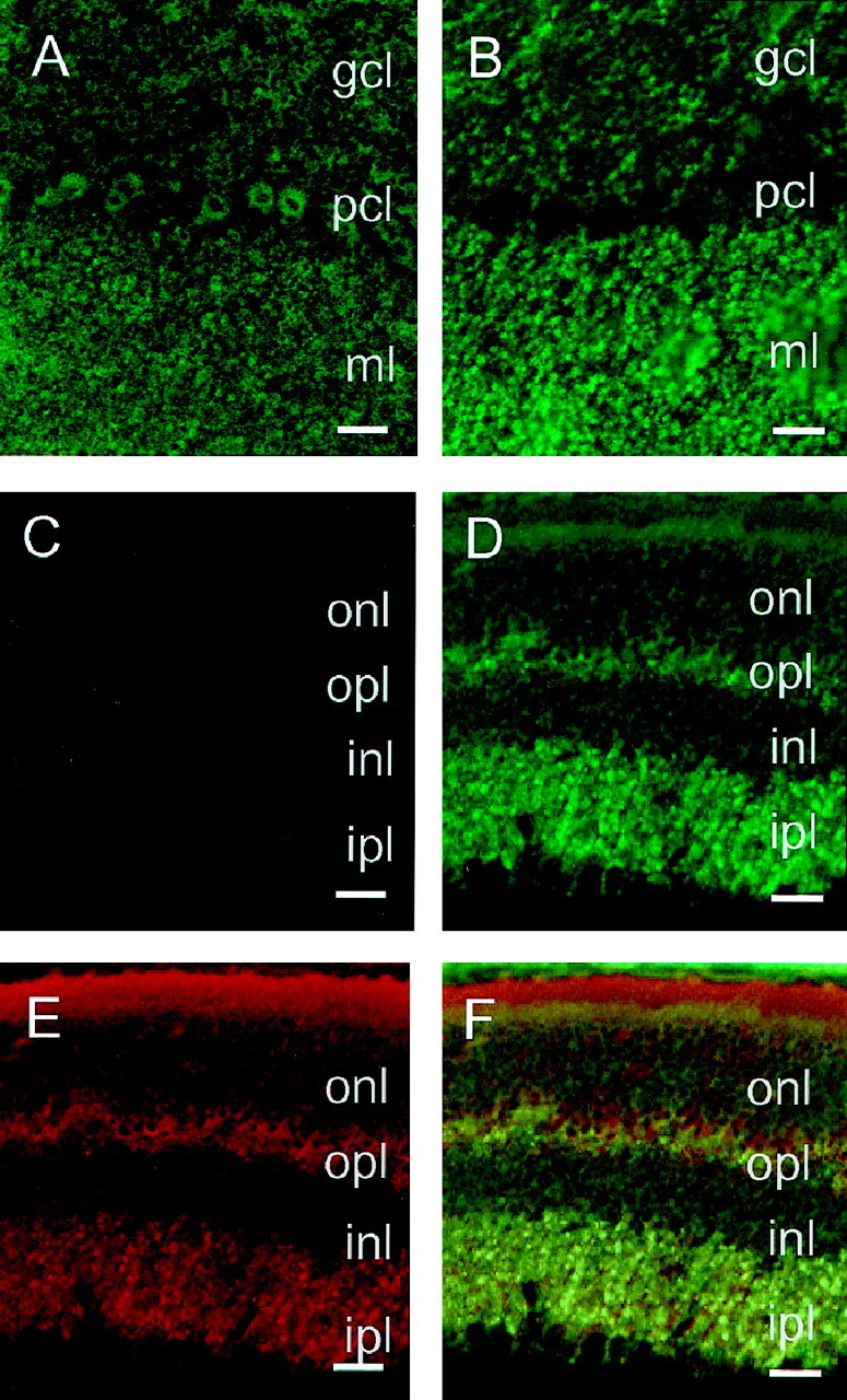
Laser scanning confocal images of PrPC in the cerebellar cortex and retina of wild-type and PrPC-overexpressing Prnp0/0-reconstituted mice. A,B, Cerebellar cortex of tg35 and tg20 animals analyzed using PrP antibody 3B5. In tg35 (A), PrPC was detected over the granule cell layer (gcl), the Purkinje cell bodies (pcl), and the molecular layer (ml). In tg20 (B), a strong PrPC expression was observed over the molecular layer and granule cell layer but not in the Purkinje cell layer.C–F, Retina of wild-type (C) and PrPC-overexpressing tg20 (D–F) mice. Using PrP antibody 3B5, no signal was detected in wild-type mice (C), whereas the tg20 retina (D) showed a strong PrPC expression in the inner plexiform layer (ipl), as well as the outer plexiform layer (opl). In tg20 mice, synaptophysin (E) was found to be coexpressed with PrPC in the outer and inner plexiform layers (F). PrPC expression is low in the outer (onl) and inner nuclear layers (inl). Scale bars: A–F, 150 μm.