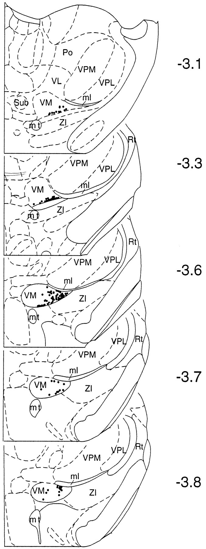
Fig. 1.

Rostrocaudal distribution of neurons recorded in the VM (n = 135) that responded to noxious cutaneous stimuli. Each neuron is presented as a dot in a schematic representation of a coronal section of the diencephalon (Paxinos and Watson, 1997). Note that most of the units recorded were located in the VMl between −3.1 and −3.8 mm with respect to bregma. ml, Medial lemniscus;mt, mammillothalamic tract; Rt, reticular thalamic nucleus; Po, posterior thalamic nucleus;Sub, submedius thalamic nucleus; VL, ventrolateral thalamic nucleus; VM, ventromedial thalamic nucleus; VPL, ventroposterolateral thalamic nucleus; VPM, ventral posteromedial thalamic nucleus;ZI, zona incerta.
