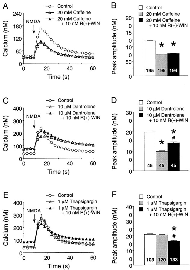
Fig. 10.
Involvement of Ca2+ stores in the cannabinoid enhancement of the Ca2+ signal to NMDA. A, B, Representative Ca2+ signals (A) and the mean ± SEM peak amplitude of the Ca2+ signals (B) evoked by NMDA in granule neurons under control conditions, in the presence of the caffeine (20 mm) and in the presence of caffeine plus 10 nmR(+)-WIN. NMDA (50 μm) was applied at thearrow by a 400 msec microperfusion pulse. Each trace inA is from a different microscopic field of neurons in the same culture dish and represents the mean ± SEM Ca2+ signal for 15 granule neurons measured individually; error bars are smaller than the corresponding symbol in many cases. The mean values in B are from all granule neurons studied in four culture sets. C,D, Representative Ca2+ signals (C) and the mean ± SEM peak amplitude of the Ca2+ signals (D) evoked by NMDA in granule neurons in studies using dantrolene (10 μm). Results are representative data from one culture set; similar results were obtained in an additional three culture sets. Studies were performed similarly to those in A andB. E, F, Representative Ca2+ signals (E) and the mean ± SEM peak amplitude of the Ca2+ signals (F) evoked by NMDA in granule neurons in studies using thapsigargin (1 μm). Results are from two culture sets; studies were performed similarly to those in A andB. Numbers in the bar graphs (B, D,F) represent the number of neurons measured for each condition. *Significant difference from control;#significant difference compared with the presence of the respective pharmacological agent (p < 0.05, ANOVA followed by post hoc analysis using Scheffé’s F test).

