
Fig. 3.
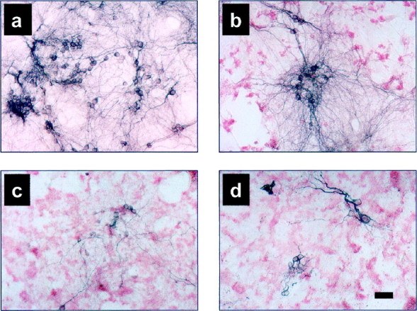
Immunocytochemistry of neurons and of Aβ in primary mixed cortical cultures exposed to various Aβ preparations. Mixed cortical cultures were exposed to medium only (a), LMW Aβ (b), PF (c), or fibrils (d) for 5 d, fixed and double-labeled for Aβ deposition (R1282) (red staining) and neurons (MAP2) (blue staining). Significant neuron loss is observed with each of the Aβ preparations (LMW Aβ, 18 μm; PF, 21 μm; fibrils, 28 μm). Scale bar, 50 μm.