
Fig. 5.
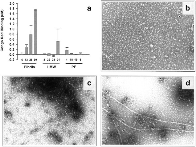
Lack of detectable fibril formation in mixed brain cultures treated for 5–6 d with LMW Aβ or PF. a,Mixed cortical cultures were exposed to LMW, PF, or preformed fibrils for 6 d, at which time the medium was removed and analyzed for fibril formation by Congo red binding. Each column is the mean of duplicate assays. Graph represents one of three identical experiments yielding similar results. b–d, Lack of fibril formation as determined by immuno-EM in 5 d conditioned media of LMW Aβ (b), PF (c), or fibril (d) preparation. PF-treated media (c) predominantly contained large electron-dense mats of distinct protofibrils. In contrast, distinct fibrils (d) could readily be detected in the media of cultures treated with preformed fibrils. Results represent one of two experiments, in both of which few or no fibrils were detected in the media of LMW Aβ and PF cultures.