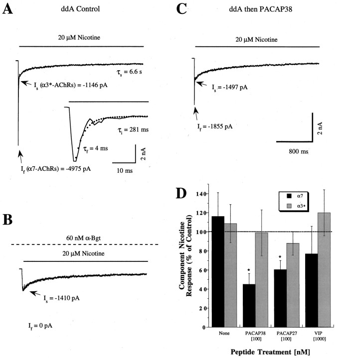
Fig. 1.
PACAP selectively inhibits α7-AChR currents when AC activity is suppressed. A–C, Representative whole-cell current records are displayed from separate E13 and E14 ciliary ganglion neurons held at −70 mV and exposed to 20 μm nicotine for ∼3 sec by fast perfusion, as indicated by the solid bar. All neurons were pretreated with 200 μm ddA for 1–7 min to inhibit AC as described in Materials and Methods. Arrows labeledIf and Isindicate peak values of the α7- and α3*-AChR current components, respectively. Calibration bars in C apply toA–C. A, Response to 20 μmnicotine application from a control neuron (Cm = 23 pF) features a rapidly activating current reaching a peak value (Ip) of −6121 pA and decaying with complex kinetics. The rapidly activating and desensitizing part of the response is shown on an expanded time scale in theinset, showing the superimposed fit to the equation in Materials and Methods (dots) and the associated values of τf and τi (τs shown above). Similar nicotine responses were obtained in the presence of 1 μm TTX and 10 μm CdCl2 (J. Margiotta, unpublished observations), indicating that they arise from nicotinic AChRs rather than from voltage-dependent Na+ or Ca2+ channels (Zhang et al., 1994). B, Nicotine response from a neuron (Cm = 28 pF) incubated with 60 nm αBgt beginning 2 hr before, and continuing throughout, the ddA pretreatment and recording (dashed bar). Note that the fast component is abolished by αBgt, indicating that it arises from α7-AChRs. C, Nicotine response from a neuron (Cm = 25 pF) treated with 100 nm PACAP38 for 6 min after the ddA pretreatment. Note that the PACAP38 treatment reduced the α7-AChR component (If), leaving the α3*-AChR component (Is) unaffected.D, Summary of the percentage change (mean ± SEM) in the α7-AChR (If/Cm) and α3*-AChR (Is/Cm) response components after treatment with neuropeptides. Each bar is based on nicotine responses like those in A andC where the individual fast and slow current components indicated by the black and gray bars, respectively, were determined as described above for the indicated peptide treatment (n = 6–19 for each concentration). Results are expressed as a percentage of the component nicotine responses obtained from the same number of control neurons tested in parallel that were treated with ddA alone. Thebars labeled none depict results from neurons treated with ddA alone, compared with untreated controls.Asterisks indicate a significant change (p < 0.05 by Student’s ttest) in the relevant current component associated with the indicated peptide treatments. Note that both PACAP38 and PACAP27 selectively reduced the α7-AChR component without affecting the α3*-AChR component.

