
Fig. 6.
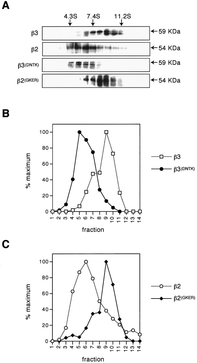
Differential sedimentation of(FLAG)β subunits on sucrose density gradients. COS cells transfected with (FLAG)β3,(FLAG)β3(DNTK), or(FLAG)β2(GKER) were subjected to sucrose density gradient fractionation 16 hr after transfection. Gradient fractions were separated by SDS-PAGE; the(FLAG)β subunits were detected by Western blotting using anti-FLAG M2 monoclonal antibody (A), and the signals were quantified using a Bio-Rad phoshorimager (B, ■, (FLAG)β3, ●,(FLAG)β3(DNTK); C, ○,(FLAG)β2, ♦,(FLAG)β2(GKER)). The data for(FLAG)β2 are taken from Gorrie et al. (1997) and represent immunoprecipitation of this protein from expressing cells after metabolic labeling with [35S]methionine. The level of β2 in each fraction was quantified using a Bio-Rad phosphorimager. Sedimentation coefficients of receptor subunits were determined by reference to the standards BSA (4.3S), aldolase (7.4S), and catalase (11.2S).