
Fig. 2.
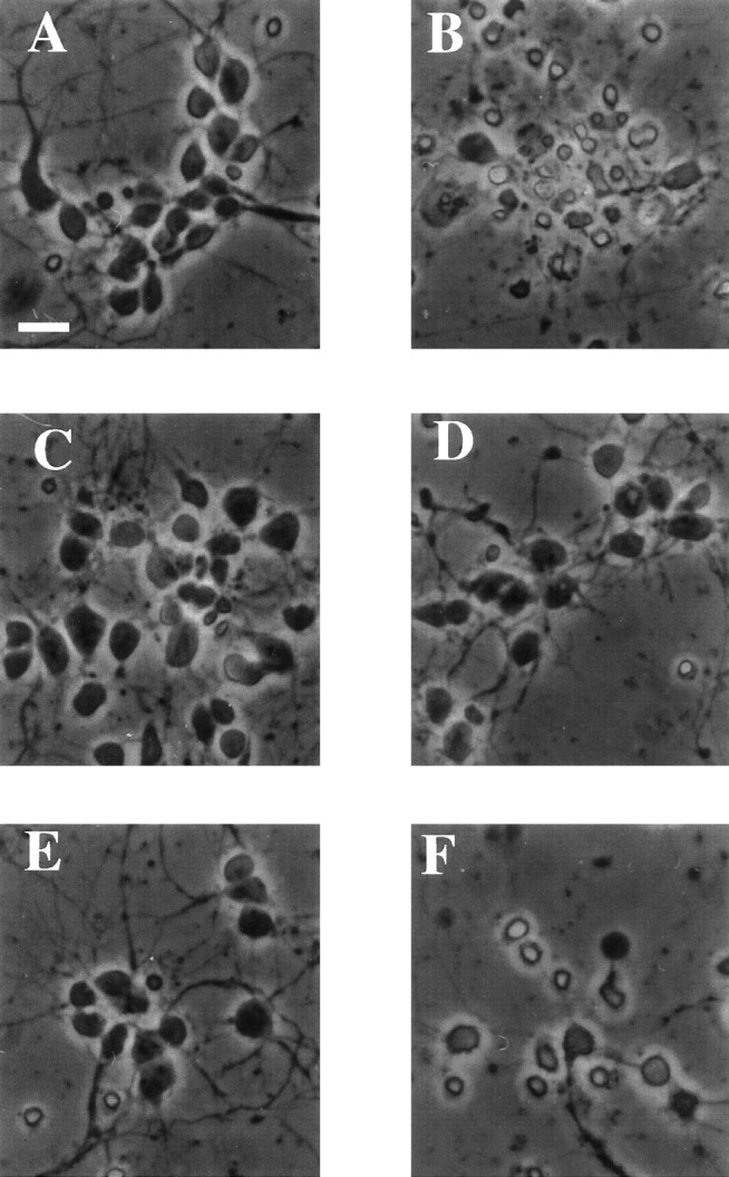
Photomicrographs of cultured E18 cortical neurons treated with camptothecin and Cdk and caspase inhibitors. Rat E18 cortical neurons were plated in poly-d-lysine-coated 24-well dishes. The following day 10 μm camptothecin, with or without 1 μm flavopiridol, or 100 μm BAF, was applied to the cells. Twelve hours later, photomicrographs were taken of cells that had been untreated (A) or treated with camptothecin alone (B) or in combination with flavopiridol (C) or BAF (D). Sister cultures were exposed for 24 hr to camptothecin and flavopiridol (E) or camptothecin and BAF (F) and photomicrographed. Scale bar, 25 μm.