
Fig. 4.
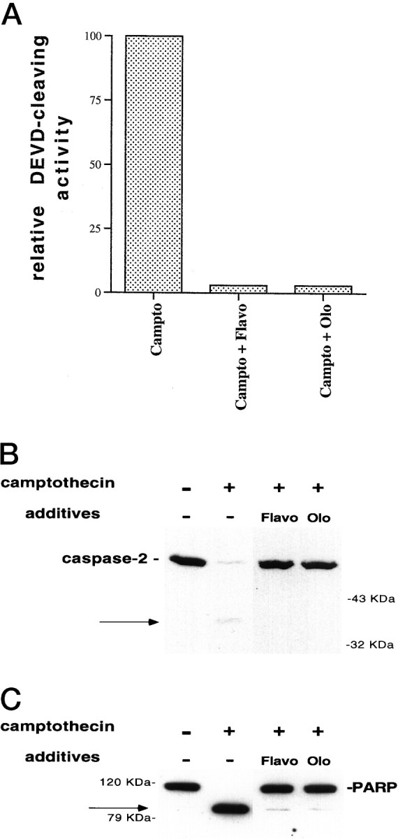
Cdk inhibitors block caspase activation in camptothecin-treated neurons. A, E18 cortical neurons were plated in poly-d-lysine-coated 35 mm dishes and the following day were treated with 10 μm camptothecin alone or together with 1 μm flavopiridol (Flavo) or 200 μm olomoucine (Olo). Ten hours later, soluble lysates were generated (10 μg) and assessed for their ability to cleave the fluorogenic substrate DEVD-AFC (see Materials and Methods). The results are representative of two separate experiments and are reported as percentage of activity of lysates treated with camptothecin alone. B, C, E18 cortical neurons were treated with camptothecin alone or in combination with flavopiridol (1 μm) or olomoucine (200 μm). Ten hours later, soluble (B) or particulate (C) neuronal lysates (50 μg) were generated as described in Materials and Methods and subjected to SDS-PAGE on a 12% (B) or 10% (C) gel. After Western immunoblotting with the anti-N-Nedd antibody (B) (1:250) or the C-2–10 monoclonal anti-PARP antibody (1:5000) (C), the bands were visualized by ECL (Amersham). The arrows indicate the 37 kDa cleavage product for caspase-2 (B) and the 85 kDa PARP cleavage product (C).