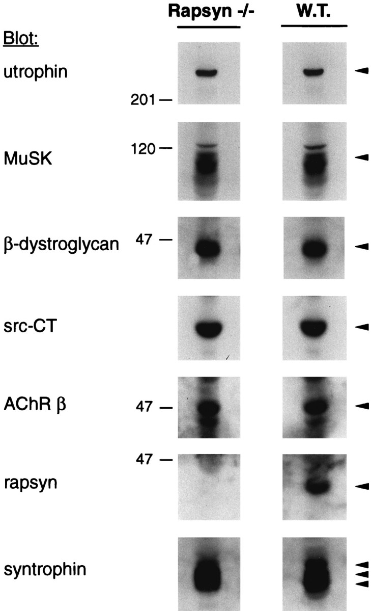
Fig. 3.

Analysis of postsynaptic proteins in rapsyn −/− and wild-type myotubes. Cells (11-7 and 12-10) were lysed in a buffer containing 1% NP-40, and protein-matches fractions, ∼1% of the extracts, were subjected to immunoblotting using utrophin-, MuSK-, or β-dystroglycan-specific antibodies. Blots were stripped and reprobed with the src-CT antibody; in a second round of stripping and reprobing, mAb 124 antibodies recognizing the AChR β subunit were used. Parallel lysates were analyzed by immunoblotting with rapsyn- or syntrophin-specific antisera. Arrowheads indicate the proteins recognized by the respective antibodies.
