
Fig. 1.
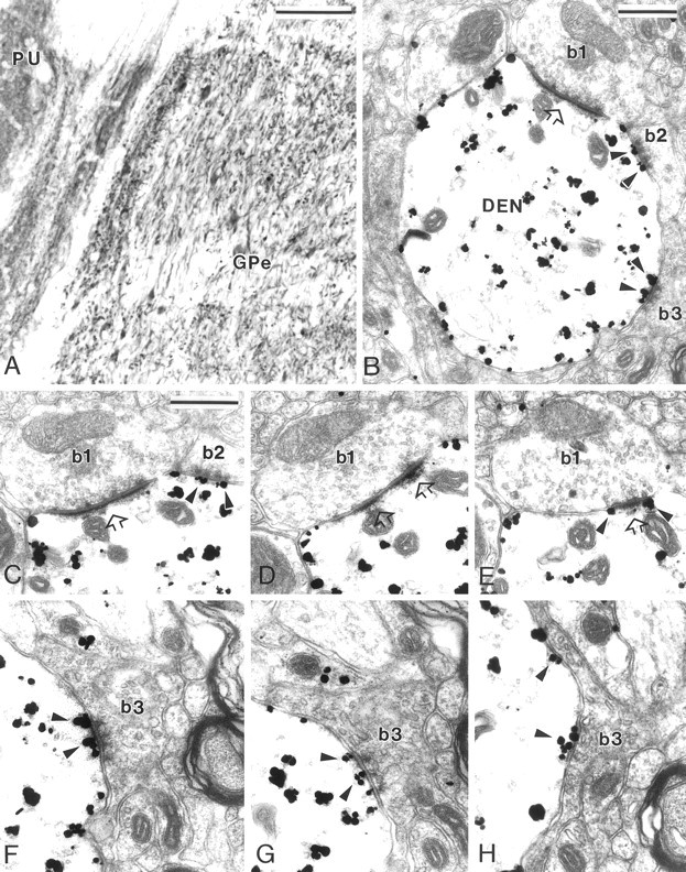
MGluR5 immunolabeling in GPe. A, Light microscopic immunoperoxidase mGluR5 labeling in GPe and putamen (PU). B, Electron micrograph of an mGluR5-positive dendrite (DEN) contacted by three boutons that display clear synaptic specializations (b1–b3). Bouton b1, which displays the ultrastructural features of a subthalamic terminal, forms an asymmetric synapse (open arrow), whereas b2 andb3, which are typical striatal boutons, form symmetric synaptic contacts. Note the gold particles located in the postsynaptic specializations of the symmetric synapses established byb2 and b3 (arrowheads).C–E, Serial ultrathin sections of b1showing the perisynaptic mGluR5 labeling (E,arrowheads) at the asymmetric synapse (open arrows). In C, the arrowheadspoint out gold particles located at the symmetric synaptic junction established by striatal-like bouton b2.F–H, Electron micrographs showing serial sections of mGluR5 labeling (arrowheads) at the postsynaptic specialization of the symmetric synapse established byb3. Note that the gold particles are found in the main body of the symmetric postsynaptic specialization in the three serial sections. Scale bars: A, 100 μm; B,C, 0.5 μm (valid forD–H).