
Fig. 2.
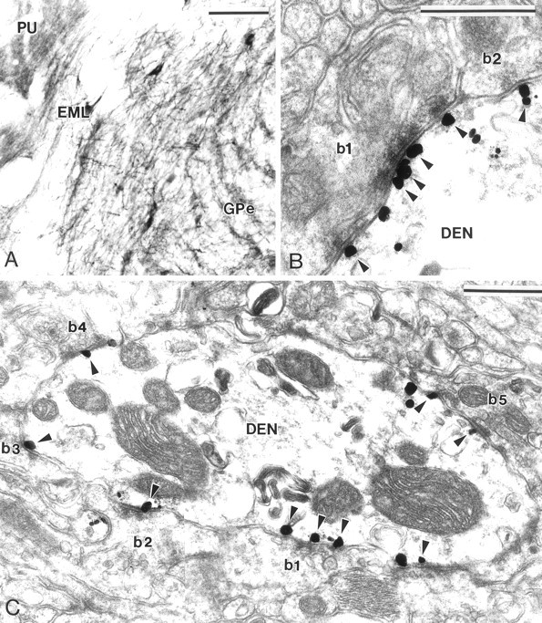
Group I mGluR immunolabeling in GPe and GPi.A, Light microscopic immunoperoxidase mGluR1a labeling in GPe and putamen (PU). Note that large neurons in the external medullary lamina (EML), which likely correspond to the cholinergic neurons of the basal nucleus of Meynert, also display strong mGluR1a immunoreactivity. B, Electron micrograph of mGluR5 immunolabeling at symmetric axodendritic synapses established by two striatal-like terminals (b1, b2). Note that gold particles aggregate in the main body of postsynaptic specializations (arrowheads).C, mGluR1a-immunoreactive dendrite (DEN) contacted by numerous striatal-like terminals (b1–b5). Most of the gold particles (arrowheads) are specifically located at the postsynaptic specialization of symmetric striatopallidal synapses. Scale bars: A, 100 μm; B, C, 0.5 μm.