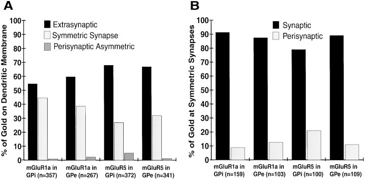
Fig. 3.
Subsynaptic distribution of group I mGluRs in GPe and GPi. A, Histogram showing the relative distribution of membrane-bound gold particle labeling for mGluR1a and mGluR5 on GPi and GPe dendrites. The total number of gold particles for each category is indicated in parentheses. A total of 125 dendrites in each pallidal segment were examined in mGluR1a- and mGluR5-immunostained sections. No gold labeling was found in the main body of the postsynaptic specializations of asymmetric synapses.B, Histogram showing the relative distribution of synaptic versus perisynaptic group I mGluRs labeling at symmetric striatopallidal synapses. For each category, the number of gold particles associated with symmetric synapses is given inparentheses.

